
Although it may seem simple and second natured, flexing and moving one’s muscles require a lot more parts than just the arm or the legs, it takes an army! In addition, it is also important to remember that our brains control every one of our movements! To go more depth with this topic, as the brain plans to move, neurons will synapse with muscles, thus creating a motor unit. Each motor unit dispenses a different amount of muscle fibers depending on the body part, for example the biceps produce a lot more muscle fibers than the pinkie toe!
 Through this neurotransmitter lab, the goal is to get a visual understanding of how the brain is connected to muscle movements through an electromyogram. To view how muscles look when they are contracting and at rest, this lab will focus on the triceps and the biceps because they are complimentary muscles; as the triceps contract (agonist contracts), the biceps relax (antagonist relaxes), and vice versa.
- To begin this lab, my group consisting of Julianna B. and Krstal M., first decided on who was going to be the test subject.
- We were given electrode pads, which we attached to Krystal’s
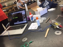 biceps and triceps and back of her hand.
 - Following the given diagram from the lab, we attached the corresponding Muscle SpikerBox leads to the electrodes on Krystal’s biceps.
- After connecting all the necessary components, we turned on the Spiker Box and connected the aux-like cable to Julianna’s computer to see the activity between the biceps and the muscle fibers/brain.
- The activity was shown when Krystal made movements such as:
- wave
- do push ups
- flex
 Although we had some difficulty retrieving clear data, from
 watching the activity from the app and the computer program, we noticed that when Krystal performed push ups and when she moved her arm, the spikes showed a lot of activity and increased in size. The left is an image of when Krystal activated her biceps, while the right is how our data appeared when she stayed still. Although the app only represented one channel, what was expected to happen was when the biceps contracted, more activity in the spikes should have appeared in comparison to the relaxing triceps, which would have resembled more of the activity on the right.
When waving, as you bring your hand towards your head, you are using your biceps. Because you are contracting the muscle, it is the agonist. When lowering the arm, the triceps are being used as they contract to allow the hand to lower, thus changing from the antagonist to the agonist.


Given the data from the lab (above), when one performs a push up, the tricep serves as the agonist for both the push up going down and up. Although the spikes decrease when going down, it is blatant that more tricep muscle fibers are in use when performing a push up.
Learn more about the lab here!
Learn more about antagonist muscles here!
